Chest wall tumours can develop in the ribs, breastbone, cartilage, muscles and other tissues of the chest wall. Treatment depends on whether the tumour is benign or malignant, its type, size, location and whether it has spread.
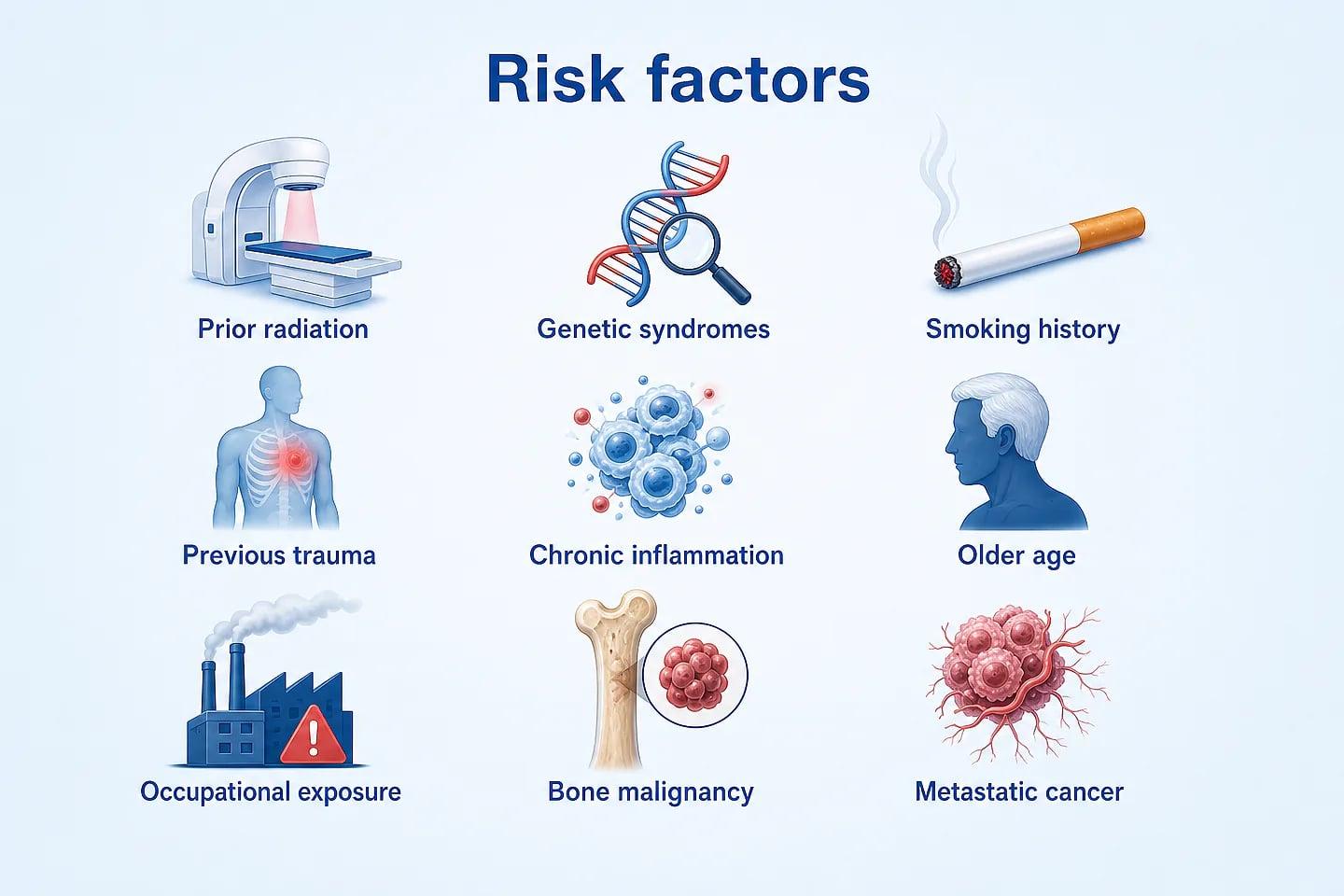
A chest wall tumour is a growth that develops in the tissues forming the outer structure of the chest.
The chest wall includes the ribs, breastbone or sternum, cartilage, muscles, fat, blood vessels and nerves. Tumours can arise from any of these structures.
Some chest wall tumours are benign and do not spread to other parts of the body. Others are malignant, including a number of bone and soft-tissue sarcomas.
A tumour may also reach the chest wall after spreading from another part of the body or by growing into the chest wall from a nearby organ.
A new or growing lump, swelling or persistent pain should be evaluated rather than assumed to be cancerous. Imaging and, when appropriate, biopsy help establish the diagnosis.
Chest wall tumours are classified according to the tissue from which they arise and whether they are benign or malignant.
A malignant tumour arising from cartilage. In the chest wall, it can develop in a rib and less commonly in the sternum.
A cancer arising from bone-forming cells. It can rarely involve the ribs or other bones of the chest wall.
A cancer that can begin in bone or soft tissue and may affect the chest wall, particularly in younger patients.
Different sarcoma types can arise from muscles, fat, blood vessels, nerves and other soft tissues of the chest wall.
Symptoms vary according to the tumour's type, size and location. Some symptoms may also occur with conditions that are not cancer.
A lump or visible growth on the chest wall may gradually increase in size and should be evaluated if it is new or changing.
Tumours involving bone or cartilage can sometimes cause persistent pain, tenderness or discomfort.
Localized swelling may occur around a chest wall tumour depending on its size and location.
A larger tumour may affect nearby structures and can sometimes contribute to breathing difficulty.
Imaging helps determine where a tumour is located and how far it extends. A biopsy may then be used to establish the exact tumour type.
Your medical history, symptoms, previous cancer treatment and physical findings are reviewed.
X-ray, CT, MRI or PET/CT may be used depending on the tumour and the information required.
A tissue sample may be obtained to determine whether the tumour is benign or malignant and identify its specific type.
If the tumour is malignant, additional tests may be required to determine its extent and whether it has spread.
Treatment depends on whether the tumour is benign or malignant, its exact type, size, location and whether it has grown into nearby structures or spread elsewhere.
Surgery is an important treatment for many malignant tumours that begin in the chest wall. The surgical approach depends on the tumour's location and extent.
When removal of a tumour creates a significant defect, reconstruction may be required to provide chest wall stability and protect structures inside the chest.
Chemotherapy may be recommended for selected tumour types, including some sarcomas, depending on the diagnosis and overall treatment strategy.
Radiation therapy may be used before or after surgery for selected tumours, or when surgery is not suitable for safely removing the tumour.
Surgery may be the main treatment for many malignant tumours that originate in the chest wall. The aim, when appropriate, is to remove the tumour completely with an adequate margin of surrounding healthy tissue.
Depending on the tumour, surgery may involve removal of part of one or more ribs, the sternum, muscle or other nearby tissue.
The exact procedure depends on the tumour's location, size, tissue of origin and relationship to surrounding structures.
If removal leaves a substantial chest wall defect, reconstruction may be considered. Depending on the case, surgeons may use mesh, plates or other materials, along with suitable tissue coverage.
Malignant chest wall tumours can require more than one treatment approach. Depending on the diagnosis, care may involve surgeons, medical oncologists, radiation oncologists, radiologists and pathologists.
Imaging and pathology help determine the tumour's location, tissue of origin, extent and relationship with nearby structures before surgery is planned.
A structured evaluation helps determine the most appropriate next step for each patient.
Existing scans, biopsy reports, medical history and previous treatment records are reviewed.
Imaging and pathology are evaluated to understand the tumour type, location and extent.
Suitable treatment approaches and their potential benefits and risks are discussed with the patient.
Surgery, systemic therapy, radiation or a combination may be planned according to the individual diagnosis.
Dr. Nitin Singhal is a surgical oncologist in Ahmedabad with experience in the surgical management of different types of cancer.
Patients with a chest wall tumour can undergo specialist evaluation to understand the diagnosis, review imaging and pathology, and determine whether surgical treatment may be appropriate.
Chest wall tumours are growths that develop in the structures forming the outer part of the chest, including the ribs, sternum, cartilage, muscles, fat and other soft tissues. They may be benign or malignant.
Malignant chest wall tumours can include different types of bone and soft-tissue sarcomas. Chondrosarcoma, osteosarcoma, Ewing sarcoma and soft-tissue sarcomas are among the tumour types that can affect the chest wall.
Diagnosis of chest wall tumours may involve a physical examination, imaging such as CT or MRI, and a biopsy to establish the exact tumour type. Additional imaging may be required when there is concern that a malignant tumour has spread.
Chest wall tumour surgery may be an important part of treatment for selected malignant tumours. Depending on the tumour, surgery may involve removal of part of the ribs, sternum, muscle or other affected tissues. Reconstruction may be required when removal creates a significant chest wall defect.
Additional treatments such as chemotherapy or radiation therapy may be recommended for certain tumour types. The appropriate treatment and sequence depend on the individual diagnosis, tumour characteristics and overall health.
Patients seeking chest wall tumour treatment in Ahmedabad can bring their imaging scans, biopsy reports and previous medical records for specialist evaluation and treatment planning.
Common questions about chest wall tumour diagnosis, surgery and treatment.
Chest wall tumours are growths that develop in structures such as the ribs, sternum, cartilage, muscles, fat, blood vessels and nerves. They can be benign or malignant.
No. Some chest wall tumours are benign and may only require observation or treatment when they cause symptoms, continue to grow or cannot be clearly identified as benign.
Malignant chest wall tumours can include chondrosarcoma, osteosarcoma, Ewing sarcoma and different types of soft-tissue sarcoma. The exact tumour type is determined through appropriate diagnostic evaluation.
Possible symptoms include a new or growing lump, chest wall pain or tenderness, swelling and, in some cases, breathing difficulty if the tumour affects nearby structures.
Evaluation can include a physical examination, chest X-ray, CT, MRI or PET/CT depending on the situation. A biopsy may be required to determine the exact type of tumour.
Surgery is a main treatment for many malignant tumours that originate in the chest wall. Whether surgery is appropriate depends on the tumour type, location, size, extent and the patient's overall health.
Reconstruction may be needed when removal of a chest wall tumour creates a significant defect. Depending on the case, materials such as mesh or plates and appropriate tissue coverage may be used to help restore chest wall stability.
Yes. Chemotherapy or radiation therapy may be appropriate for certain chest wall tumours. Their use depends on the tumour type, stage and overall treatment plan.
Treatment depends on the exact tumour type, tumour size and location, grade and stage, involvement of nearby structures, whether the tumour has spread and the patient's overall health.
A new or growing chest wall lump, persistent swelling, or chest wall pain that does not resolve should be medically evaluated. A specialist can determine whether imaging or other investigations are appropriate.
Bring your scans, biopsy reports and previous medical records for specialist evaluation and an individualized discussion of available treatment options.
Book a ConsultationExplore specialised cancer treatment and surgical oncology services, organised by area of expertise.
Advanced surgical approaches for selected complex cancers.
Specialised surgical oncology care for women's cancers.
Specialised treatment and surgical care for ovarian cancer.
Comprehensive care for selected gynaecological cancers.
Individualised cervical cancer evaluation and surgical care.
Surgical and multidisciplinary care for uterine cancers.
Specialised surgical care for gastrointestinal cancers.
Cancer care for the urinary system and male reproductive organs.
Comprehensive surgical care for urological cancers.
Specialised surgical treatment for kidney cancer.
Personalised surgical care for prostate cancer.
Evaluation and surgical treatment of testicular cancer.
Diagnosis and surgical management of bladder cancer.
Specialised surgical oncology care for head and neck cancers.
Specialised surgical care for breast cancer.
Specialised cancer care for children and young patients.

Dr. Nitin Singhal has been recognised for his contribution to cancer care and surgical oncology, reflecting his commitment to advanced surgical techniques, clinical excellence and patient-centred care.
Surgical oncology training from Tata Memorial Hospital, Mumbai.
Expertise across complex and multidisciplinary cancer surgery.
Honoured for excellence in healthcare and surgical oncology.
Focus on evidence-based and personalised cancer treatment.
Recognition that reflects a continued commitment to excellence in cancer care.