Comprehensive evaluation and treatment planning for vaginal cancer, with the approach selected according to tumour location, size, stage, lymph node involvement and the patient's overall health.
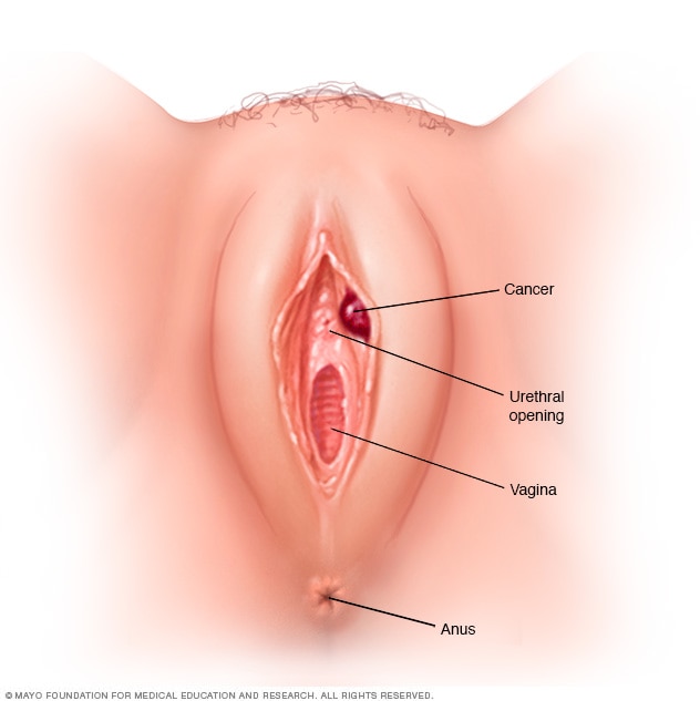
Vaginal cancer develops in the cells of the vagina, the muscular canal extending from the cervix to the outside of the body.
Primary vaginal cancer is uncommon. Squamous cell carcinoma is the most common type, while adenocarcinoma and several rarer tumour types can also occur.
Treatment depends on several factors, including the location and size of the tumour, stage of disease, lymph node involvement, previous pelvic treatment and the relationship of the tumour to nearby organs such as the bladder and rectum.
Primary vaginal cancer is uncommon and requires careful assessment to distinguish it from cancers that have spread to the vagina from another site.
The location of a vaginal tumour can influence lymphatic drainage, surgical options and radiation planning.
The upper portion lies close to the cervix and uterus and has lymphatic drainage that may involve the pelvic nodes.
Tumours in the middle portion are assessed according to their depth, size and relationship to surrounding structures.
Tumours involving the lower vagina may have lymphatic drainage toward the inguinal and pelvic lymph nodes.
The vagina lies close to the bladder, urethra and rectum, which can influence both surgery and radiation planning.
Squamous cell carcinoma accounts for the majority of primary vaginal cancers and develops from the squamous cells lining the vagina.
Adenocarcinoma develops from glandular cells and represents a smaller proportion of primary vaginal cancers.
Melanoma can rarely arise in the vagina and requires specialised pathological and oncological assessment.
Sarcomas, neuroendocrine tumours and other rare malignancies can occasionally arise in vaginal tissue.
Certain high-risk types of human papillomavirus are associated with many vaginal cancers.
HPV is a common infection, and most infections do not lead to cancer. Persistent infection with certain high-risk HPV types can cause cellular changes that may eventually become cancerous.
HPV-related vaginal precancer is known as vaginal intraepithelial neoplasia, or VaIN. Some vaginal precancerous lesions can be monitored or treated before invasive cancer develops.
HPV vaccination and appropriate cervical and gynecologic screening can help reduce the risk of HPV-related cancers.
NCI reports that HPV infection causes a substantial proportion of vaginal cancers. HPV vaccination may help reduce the risk.
Early vaginal cancer may not cause noticeable symptoms. Possible symptoms include:
Bleeding after sexual intercourse, between periods or after menopause can require medical evaluation.
Persistent watery, bloody or unusual vaginal discharge may occur.
Pelvic discomfort or pain can occur, particularly with more extensive disease.
Persistent pain during sexual intercourse can be a symptom that warrants evaluation.
A tumour close to the urethra or bladder can sometimes affect urinary function.
More advanced disease involving nearby structures can produce bowel or rectal symptoms.
VaIN describes abnormal cells in the vaginal lining that have not invaded deeper tissues.
Low-grade lesions may regress naturally and can often be managed with careful observation.
Intermediate-grade changes may be monitored or treated depending on the lesion and individual circumstances.
High-grade lesions are considered at increased risk of progression and generally require active treatment.
Selected lesions may be treated with laser therapy, wide local excision, vaginectomy, topical treatment or internal radiation.
Diagnosis generally begins with a pelvic examination and careful assessment of any abnormal vaginal area.
A biopsy is required to establish whether abnormal tissue represents cancer and to determine the tumour type.
Imaging studies may then be used to evaluate the size and extent of the tumour and to assess nearby lymph nodes or distant disease when appropriate.
Tumour size, location, local extension and lymph node involvement help determine the most appropriate treatment approach.
Examination can identify visible or palpable abnormalities in the vagina and surrounding structures.
Magnified examination can help identify suspicious vaginal areas that require tissue sampling.
Tissue examination establishes the tumour type and provides important pathological information.
MRI, CT or other imaging may be used to assess local disease, lymph nodes and possible distant spread.
For selected stage I cancers, surgery, radiation or a combination may be considered depending on tumour location and characteristics.
A localized vaginal tumour may be removed together with a margin of surrounding tissue when an appropriate surgical approach is possible.
Removal of part or all of the vagina may be considered for selected tumours depending on their size, depth and anatomical location.
The extent of surgery depends strongly on the location and depth of the tumour and its relationship to nearby organs.
Selected localized lesions may be removed with a surrounding margin of healthy tissue.
A portion of the vagina containing the tumour may be removed when an appropriate margin can be achieved.
More extensive vaginal disease may require removal of the entire vagina in selected patients.
Extremely extensive recurrent or locally advanced disease may require pelvic exenteration in carefully selected patients.
The location of the primary tumour influences lymphatic drainage and can affect both surgery and radiation planning.
Tumours in the upper vagina may have lymphatic drainage toward the pelvic lymph nodes and may lie close to the cervix and uterus.
Tumours involving the lower third of the vagina may have lymphatic drainage toward the inguinal as well as pelvic lymph nodes.
Lymphatic drainage differs according to tumour location, making regional lymph node assessment an important part of treatment planning in selected patients.
Tumours in the upper and middle vagina may drain toward pelvic lymph nodes.
Tumours involving the lower vagina may drain toward the inguinal lymph nodes.
Lymph node removal may be considered during surgery depending on tumour location and stage.
Regional lymph nodes may be included in radiation treatment when clinically appropriate.
Radiation is an important treatment for many vaginal cancers, particularly when surgery is not appropriate or when disease is more locally advanced.
Radiation is delivered from outside the body to the tumour and relevant regional lymph node areas.
A radioactive source is positioned inside or close to the tumour to deliver a concentrated dose of radiation.
External radiation may be combined with brachytherapy depending on tumour location, size and stage.
Chemotherapy may be administered with radiation in selected locally advanced vaginal cancers.
More extensive disease may involve surrounding tissues or regional lymph nodes and often requires multidisciplinary treatment planning.
Radiation is commonly used for stage II, III and selected stage IVA vaginal cancers.
Concurrent chemotherapy and radiation may be considered in selected patients.
Radical vaginectomy or pelvic exenteration may be considered in selected cases where surgery is appropriate.
Surgical oncology, radiation oncology and medical oncology may coordinate the treatment strategy.
Advanced vaginal cancer may extend to nearby structures or spread to distant organs. Treatment is selected according to the extent of disease and the patient's previous treatment.
Radiation therapy can be used to control local disease or relieve symptoms. Chemotherapy may also be considered, particularly when disease cannot be managed with local treatment alone.
Because vaginal cancer is rare, evidence for systemic treatment in advanced disease is limited and treatment may be based partly on approaches used for cervical cancer.
Treatment for advanced or recurrent disease depends on previous radiation or surgery, the location of recurrence and the extent of disease.
Recurrent disease requires reassessment of the location of recurrence and all previous treatments.
Selected local recurrences may be evaluated for surgery or radiation depending on previous treatment.
Extensive pelvic recurrence may require assessment for complex surgical treatment in carefully selected patients.
Previous pelvic radiation is an important factor when considering additional radiation or surgery.
Widespread recurrent disease may require systemic treatment or consideration of clinical trials.
When extensive surgery is required, reconstructive options may be discussed according to the patient's anatomy, treatment goals and overall health.
Reconstruction may be considered after extensive removal of vaginal tissue in selected patients.
Skin grafting can sometimes be used as part of reconstruction after partial or total vaginectomy.
Surgery close to the bladder or urethra requires careful planning to protect or reconstruct urinary function when possible.
Tumours close to the rectum require careful assessment because treatment can affect bowel function.
Several anatomical and clinical factors are considered before selecting surgery, radiation or combined treatment.
Establish the tumour type through examination and biopsy.
Determine its location, size, depth and relationship to nearby structures.
Consider pelvic and/or inguinal lymphatic drainage according to tumour location.
Determine whether surgery, radiation, chemoradiation or a combined approach is appropriate.

Dr. Nitin Singhal is a surgical oncologist in Ahmedabad with experience in complex cancer surgery and multidisciplinary cancer care.
Vaginal cancer requires careful assessment of tumour location, depth, stage and its relationship with nearby pelvic organs. Surgical options are considered according to the individual disease characteristics and overall treatment plan.
Vaginal cancer is a rare cancer that develops in the vagina. Most primary vaginal cancers are squamous cell carcinomas, while adenocarcinoma and several rarer tumour types can also occur.
Persistent infection with certain high-risk HPV types is associated with many vaginal cancers. Vaginal intraepithelial neoplasia (VaIN) represents precancerous changes in the vaginal lining and can sometimes be treated before invasive cancer develops.
Diagnosis generally involves pelvic examination, biopsy and imaging when appropriate. Tumour location is particularly important because upper and lower vaginal tumours can have different lymphatic drainage patterns.
Treatment may include wide local excision, partial or total vaginectomy, radiation therapy or combinations of treatment. For locally advanced disease, radiation with or without chemotherapy is commonly considered, while extensive recurrent disease may require highly specialised surgical assessment.
If you have been diagnosed with vaginal cancer or have an abnormal vaginal lesion requiring evaluation, a specialist consultation can help clarify the diagnosis, stage and available treatment options.
Common questions about vaginal cancer symptoms, diagnosis and treatment.
Get a personalised evaluation to understand your diagnosis, tumour location, cancer stage and available surgical and multidisciplinary treatment options.

Dr. Nitin Singhal has been recognised for his contribution to cancer care and surgical oncology, reflecting his commitment to advanced surgical techniques, clinical excellence and patient-centred care.
Surgical oncology training from Tata Memorial Hospital, Mumbai.
Expertise across complex and multidisciplinary cancer surgery.
Honoured for excellence in healthcare and surgical oncology.
Focus on evidence-based and personalised cancer treatment.
Recognition that reflects a continued commitment to excellence in cancer care.